Thursday, July 1, 2021

Embryo selection aimed at ensuring intelligence of children

Embryo selection aimed at ensuring intelligence of children

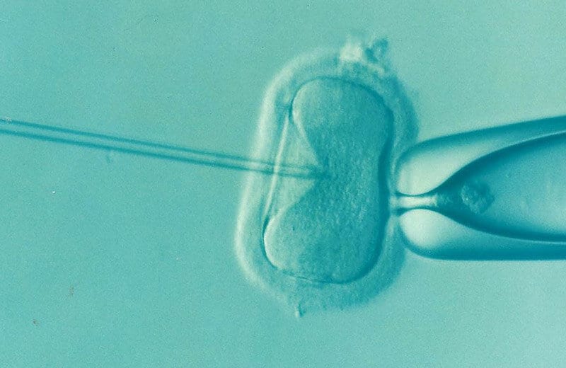
A special report published in the New England Journal of Medicine raises serious questions about the benefits, risks and ethics of a new service — which the authors call “embryo selection based on polygenic scores,” or ESPS. The service allows in vitro fertilization patients to select embryos with the goal of choosing healthier and even smarter children.

In the report “Problems with Using Polygenic Scores to Select Embryos,” the multinational team of researchers describes the limitations of the service. They also warn that patients, and even in vitro fertilization clinicians, may think that the service is more effective and less risky than it is.

The authors highlight that since the same gene often influences many different traits, selecting for one trait can lead to the unintentional selection of adverse traits. They also warn that using the service has the potential to alter population demographics, exacerbate socioeconomic inequalities and devalue certain traits.

If ESPS continues to be available to IVF patients, the researchers call on the U.S. Federal Trade Commission to develop and enforce standards for responsible communication about the service. The authors also call for a society-wide conversation about the ethical use of the technology and whether it should be regulated.

An imperfect way to ensure healthy children

Polygenic scores are predictions of individual health and other outcomes derived from studies of the genome. In adults, polygenic scores have been shown to partially predict those outcomes. However, their predictive power is significantly reduced when comparing embryos to one another, the report authors explain.

“Polygenic scores are already only weak predictors for most individual adult outcomes, especially for social and behavioral traits, and there are several factors that lower their predictive power even more in the context of embryo selection,” said Patrick Turley, assistant professor (research) of economics at the USC Dornsife College of Letters, Arts and Sciences and co-first author of the paper. “Polygenic scores are designed to work in a different setting than an IVF clinic. These weak predictors will perform even worse when used to select embryos.”

Turley, who is also director of the Behavioral and Health Genomics Center at the USC Dornsife Center for Economic and Social Research, and his colleagues on the study examined if ESPS is any more effective at ensuring future health than random selection of embryos. To do so, they modeled the expected difference in a future individual’s risk for several diseases, comparing the use of ESPS to select an embryo versus choosing one viable embryo at random from among 10. They found that, in most cases, the reduction of disease risk the service offers is very small and highly uncertain.

Multiple companies are now working with IVF clinics to offer the service to patients who want to select an embryo with a lower chance of growing up to develop diabetes, cancer, heart disease, Alzheimer’s disease, inflammatory bowel disease and schizophrenia. One company also offers the service for the purpose of selecting embryos according to their predicted educational attainment, household income and cognitive ability. The founder of another company has not ruled out someday offering ESPS in some countries to choose skin color or above-average mental ability.

Drawbacks to the selection service 

For ESPS to work, polygenic scores need to give at least moderately accurate predictions of whether the selected embryos will end up developing a certain trait or not. The studies that generate the polygenic scores sometimes suggest moderate or even large differences in actual outcomes between people with high versus low scores, but those differences are based on a sample of people from different families. As Turley and colleagues note, ESPS usually involves comparing members of the same family, which significantly lowers its predictive power.

Additionally, the studies that produce polygenic scores involve people with similar ancestries and mostly European ancestries. As a result, most polygenic scores constructed today will be less predictive for people of other ancestries.

Finally, assessments of the predictive power of polygenic scores typically assume very similar environments for the generation enrolled in the original study and the generation that will be born as a result of ESPS. But by the time an embryo selected by the service is an adult, the person may face a very different environment.

Widespread use of ESPS raises other risks, as well. For instance, the researchers warn that use of the service could exacerbate existing health and other disparities, as it is largely only accessible to the relatively wealthy and it currently works best among those with European ancestries. It might also amplify prejudice and discrimination by signaling that existing people with traits that parents select against are less valuable.

“Some countries have authorities that decide which traits embryos can be tested for,” said Michelle N. Meyer, assistant professor of bioethics and a legal scholar at Geisinger Health System and co-first author of the special report. “But in the U.S., there is a strong legal and ethical tradition of viewing reproductive decisions as matters of private individual choice. In the short term, the FTC should help establish what counts as adequate evidence to support claims about the expected benefits of ESPS and what counts as adequate information disclosure in this context.”

The researchers also call for professional medical societies to develop policies and guidance and for companies themselves to demonstrate that the information they provide to diverse customers is complete, accurate and well-understood.

They also say there needs to be a society-wide conversation about whether existing legal frameworks can adequately ensure accurate information about ESPS, and if limits on the use of the service should be adopted.

“Many individual reproductive decisions, aggregated over generations, can have profound societal consequences,” said Daniel J. Benjamin, corresponding author and a professor at the UCLA Anderson School of Management and David Geffen School of Medicine. “Collectively, these decisions could alter population demographics, exacerbate inequalities and devalue traits that are selected against.”

About the study

In addition to Turley, Meyer and Benjamin, authors on the study include Nancy Wang of the National Bureau of Economic Research; David Cesarini of New York University; Evelynn Hammonds and David Laibson of Harvard University; Alicia R. Martin, Ben M. Neale and Heidi L. Rehm of the Broad Institute of Harvard and MIT; Louise Wilkins-Haug of Harvard Medical School; Steven Hyman, M.D., Harvard University and Broad Institute of Harvard and MIT; and Peter M. Visscher of the University of Queensland, Australia.

The research was supported by the National Institutes of Health, Open Philanthropy, the Ragnar Söderberg Foundation, the Pershing Square Fund for Research on the Foundations of Human Behavior, the Robert Wood Johnson Foundation, the Russell Sage Foundation, the JPB Foundation, the National Health and Medical Research Council, the Stanley Family Foundation and the Australian Research Council.


from ScienceBlog.com https://ift.tt/3hsHzSe

Prenatal exposure to THC, CBD affects offspring’s responsiveness to Prozac

Prenatal exposure to THC, CBD affects offspring's responsiveness to Prozac

Scientists at Indiana University have found that significant amounts of the two main components of cannabis, THC and CBD, enter the embryonic brain of mice in utero and impair the mice’s ability as adults to respond to fluoxetine, a drug commonly used to treat anxiety and depression and known by the brand name Prozac.

The study suggests that when the developing brain is exposed to THC or CBD, normal interactions between endocannabinoid and serotonin signaling may be diminished as they become adults.

“Hemp-derived CBD is a legal substance in the U.S., and we are in a time of increasing state-level legalization of cannabis. Therefore, use of cannabis components have increased across most levels of society, including among pregnant women,” said Hui-Chen Lu, author of the study, director of the Linda and Jack Gill Center and a professor in the Department of Psychological and Brain Sciences in the IU Bloomington College of Arts and Sciences. “The study marks the beginning of an effort to understand the effects of THC and CBD on the endogenous cannabinoid system in the developing brain and body.”

The study was published in Cannabis and Cannabinoid Research and will be a part of the upcoming 2021 Gill Symposium, which will focus exclusively on the topic of cannabis.

Researchers studied four groups of pregnant mice. Some received daily moderate doses of either THC, CBD, or a combination of equal parts THC and CBD; a control group was given placebo injections throughout pregnancy. Using mass spectrometry, IU psychological and brain sciences professor Heather Bradshaw tested embryos and found that CBD and THC both could reach the embryonic brain, determining that the drug was making it past the placenta.

“The surprising part is that maternal exposure to CBD alone — a drug that is often considered as safe and harmless and is a popular ‘natural’ therapy for morning sickness — resulted in a lasting impact on adult mice offspring,” Lu said. “Both prenatal THC and CBD exposure impaired the adult’s ability to respond to fluoxetine. The results suggest taking a cautious approach to using CBD during pregnancy.”

There is some evidence for CBD’s effectiveness in treating chronic pain and anxiety, though the only FDA-approved indication for CBD to date is the treatment of severe seizure disorders.

“We still know very little about the effects of CBD on the developing brain,” Lu said.

The new paper is one of the first studies to see the potential negative impact of CBD on the developing brain and later behaviors.

Study co-author Ken Mackie, Gill Chair of Neuroscience at IU Bloomington, said researchers know that prenatal cannabis exposure may increase the risk for anxiety and depression, so it is important to evaluate the response to a class of drug used to treat anxiety and depression.

While many of the tests reflected normal mouse behaviors, one test — to determine their response to stress — stood out strongly as atypical. The mice in all groups responded normally to a stressful situation. As expected, fluoxetine increased stress resilience in mice whose mothers had received the placebo. However, the drug was ineffective in mice whose mothers had received THC, CBD or their combination.

Fluoxetine works by increasing the amount of serotonin available at brain synapses, an effect known to require the endocannabinoid system. This internal system of receptors, enzymes and molecules both mediates the effects of cannabis and plays a role in regulating various bodily systems, such as appetite, mood, stress and chronic pain.

To test if maternal exposure to THC and/or CBD impaired endocannabinoid signaling in the adult offspring, the researchers tested whether giving a drug to boost the endocannabinoid system would restore fluoxetine’s effectiveness. They found that enhancing the endocannabinoid system restored normal fluoxetine responses in mice that had received THC or CBD while their brains were developing.

Additional authors on the study are Izaque de Sousa Maciel, Gabriel de Abreu, Clare Johnson and Rida Bonday, all in the Gill Center and the IU Department of Psychological and Brain Sciences.

The study was funded by the IU Responding to the Addictions Crisis Grand Challenge, startup funds from the IU Bloomington College of Arts and Sciences and the Gill Endowment fund, as well as by the National Institute on Drug Abuse, which is part of the National Institutes of Health.


from ScienceBlog.com https://ift.tt/3ygcs30

Pacemaker-Like Devices Could Restore Lost Oral Function

Pacemaker-Like Devices Could Restore Lost Oral Function

Even the mundane act of swallowing requires a well-coordinated dance of more than 30 muscles of the mouth. The loss of function of even one of these due to disease or injury can be debilitating. For these people, nerve stimulation offers a ray of hope to regain some of their lost oral function.

In a new study, researchers at Texas A&M University have delineated the minimum size of electrical currents needed to provide sensation in different parts of the mouth. The researchers said their study is a first but vital step toward building electrical stimulation implants that can restore essential intraoral functions that are lost due to nerve or brain damage.

The results of the study are published in the journal Institute of Electric and Electronics Engineers’ (IEEE) Transactions on Biomedical Engineering.

Many essential bodily functions are coordinated by the nervous system via sensorimotor feedback loops. As the name suggests, these neural circuits involve the brain interpreting incoming signals from sensory nerves and then commanding the motor nerves to execute a certain movement. For example, sensorimotor loops play a vital role in voluntary functions, like walking or holding an object, and involuntary movements, like sneezing or blinking.

Within the mouth, also referred to as the intraoral cavity, there is a rich supply of both sensory and motor nerves. In particular, sensorimotor nerves in the soft palate and tongue coordinate several intraoral movements related to swallowing, speech and respiration. And so, damage to either the sensory or motor nerve fibers due to neurotrauma or disease can compromise these essential functions, reducing the quality of life of those afflicted.

Electrical nerve stimulation might help jumpstart the nerves into action, much like how a pacemaker can electrically stimulate nerves in the heart, causing the heart muscle to contract. But unlike a pacemaker, the details on the frequency and amplitude of the electrical currents needed for proper stimulation of different parts of the mouth have not been investigated.

“Electrical stimulation can modulate nerve currents or action potentials, which are the mode of communication to and from the brain,” said Hangue Park, assistant professor in the Department of Electrical and Computer Engineering. “And so, electrical stimulation should be carefully applied, because if not, then it might cause undesirable effects, or it might not stimulate anything at all.”

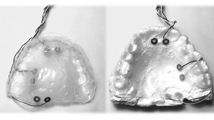
To investigate the minimum stimulation currents needed, Park and his team inserted tiny metal electrodes into a standard dental retainer. These electrodes were positioned in subjects to stimulate either their soft palate or the side and tip of the tongue, which receive a rich supply of sensory nerves. For each of these locations, the researchers slowly changed the amplitude of the stimulation current, keeping the frequency fixed. Then, subjects were asked to report when they just began feeling a sensation and when the sensation was uncomfortable. Next, they repeated the same experiment for a higher frequency of current.

After compiling their data, the team determined the average perception and discomfort thresholds for the tongue and soft palate. In addition, they produced an equivalent circuit of the intraoral cavity to duplicate the electrical properties of that area. This circuit, the researchers said, can help to further study the effects of electrical stimulation offline without requiring human subjects.

The researchers noted that their next steps would be to electrically stimulate the intraoral region and investigate how these simulations change chewing, swallowing and other behaviors.

“Sensorimotor systems can be extremely vulnerable to damage due to neural defects, aging and neurodegenerative diseases,” Park said. “In this study, we have begun to lay the groundwork for electrically stimulating parts of the mouth that control involuntary and voluntary movements. Our work is a seminal study and it is important so that we can, in the near future, help people that face enormous challenges doing everyday tasks that we take for granted.”

Other contributors to this study include Beomhee Park from the electrical and computer engineering department and Saurabh Biswas, associate professor of practice from the Department of Biomedical Engineering.

This research was funded by the Institute for Rehabilitation and Research Foundation.


from ScienceBlog.com https://ift.tt/3hnY4PI

More fish fats, less vegetable oils can reduce migraine headaches

More fish fats, less vegetable oils can reduce migraine headaches

A diet higher in fatty fish helped frequent migraine sufferers reduce their monthly number of headaches and intensity of pain compared to participants on a diet higher in vegetable-based fats and oils, according to a new study. The findings by a team of researchers from the National Institute on Aging (NIA) and the National Institute on Alcohol Abuse and Alcoholism (NIAAA), parts of the National Institutes of Health; and the University of North Carolina (UNC) at Chapel Hill, were published in the July 3 issue of The BMJ.

This study of 182 adults with frequent migraines expanded on the team’s previous work on the impact of linoleic acid and chronic pain. Linoleic acid is a polyunsaturated fatty acid commonly derived in the American diet from corn, soybean, and other similar oils, as well as some nuts and seeds. The team’s previous smaller studies explored if linoleic acid inflamed migraine-related pain processing tissues and pathways in the trigeminal nerve, the largest and most complex of the body’s 12 cranial nerves. They found that a diet lower in linoleic acid and higher in levels of omega-3 fatty acids (like those found in fish and shellfish) could soothe this pain pathway inflammation.

In a 16-week dietary intervention, participants were randomly assigned to one of three healthy diet plans. Participants all received meal kits that included fish, vegetables, hummus, salads, and breakfast items. One group received meals that had high levels of fatty fish or oils from fatty fish and lowered linoleic acid. A second group received meals that had high levels of fatty fish and higher linoleic acid. The third group received meals with high linoleic acid and lower levels of fatty fish to mimic average U.S. intakes.

During the intervention period, participants monitored their number of migraine days, duration, and intensity, along with how their headaches affected their abilities to function at work, school, and in their social lives, and how often they needed to take pain medications. When the study began, participants averaged more than 16 headache days per month, over five hours of migraine pain per headache day, and had baseline scores showing a severe impact on quality of life despite using multiple headache medications.

The diet lower in vegetable oil and higher in fatty fish produced between 30% and 40% reductions in total headache hours per day, severe headache hours per day, and overall headache days per month compared to the control group. Blood samples from this group of participants also had lower levels of pain-related lipids. Despite the reduction in headache frequency and pain, these same participants reported only minor improvements in migraine-related overall quality of life compared to other groups in the study.

Migraine, a neurological disease, ranks among the most common causes of chronic pain, lost work time, and lowered quality of life. More than 4 million people worldwide have chronic migraine (at least 15 migraine days per month) and over 90% of sufferers are unable to work or function normally during an attack, which can last anywhere from four hours to three days. Women between the ages of 18 and 44 are especially prone to migraines, and an estimated 18% of all American women are affected. Current medications for migraine usually offer only partial relief and can have negative side effects including sedation, and the possibility of dependence or addiction.

“This research found intriguing evidence that dietary changes have potential for improving a very debilitating chronic pain condition like migraine without the related downsides of often prescribed medications,” said Luigi Ferrucci, M.D., Ph.D., scientific director of NIA.

The NIH team was led by Chris Ramsden, a clinical investigator in the NIA and NIAAA intramural research programs, and UNC adjunct faculty member. Ramsden and his team specialize in the study of lipids — fatty acid compounds found in many natural oils — and their role in aging, especially chronic pain and neurodegenerative conditions. The UNC team was led by Doug Mann, M.D., of the Department of Neurology, and Kim Faurot, Ph.D., of the Program on Integrative Medicine. Meal plans were designed by Beth MacIntosh, M.P.H., of UNC Healthcare’s Department of Nutrition and Food Services.

“Changes in diet could offer some relief for the millions of Americans who suffer from migraine pain,” said Ramsden. “It’s further evidence that the foods we eat can influence pain pathways.”

The researchers noted that these findings serve as validation that diet-based interventions increasing omega-3 fats while reducing linoleic acid sources show better promise for helping people with migraines reduce the number and impact of headache days than fish-oil based supplements, while reducing the need for pain medications. They hope to continue to expand this work to study effects of diet on other chronic pain conditions.

This study was supported by the NIH NIA and NIAAA intramural research programs; and NIH grants including 1R01AT007813–01A1, T32 AT003378, DK056350, and UL1TR002489.


from ScienceBlog.com https://ift.tt/3duGtEj

Eating disorder behaviors alter reward response in the brain

Eating disorder behaviors alter reward response in the brain

Researchers have found that eating disorder behaviors, such as binge-eating, alter the brain’s reward response process and food intake control circuitry, which can reinforce these behaviors. Understanding how eating disorder behaviors and neurobiology interact can shed light on why these disorders often become chronic and could aid in the future development of treatments. The study, published in JAMA Psychiatry, was supported by the National Institutes of Health.

“This work is significant because it links biological and behavioral factors that interact to adversely impact eating behaviors,” said Janani Prabhakar, Ph.D., of the Division of Translational Research at the National Institute of Mental Health, part of NIH. “It deepens our knowledge about the underlying biological causes of behavioral symptom presentation related to eating disorders and will give researchers and clinicians better information about how, when, and with whom to intervene.”

Eating disorders are serious mental illnesses that can lead to severe complications, including death. Common eating disorders include anorexia nervosa, bulimia nervosa, and binge-eating disorder. Behaviors associated with eating disorders can vary in type and severity and include actions such as binge-eating, purging, and restricting food intake.

In this study, Guido Frank, M.D.(link is external), at the University of California San Diego, and colleagues wanted to see how behaviors across the eating disorder spectrum affect reward response in the brain, how changes in reward response alter food intake control circuitry, and if these changes reinforce eating disorder behaviors. The study enrolled 197 women with different eating disorders (including anorexia nervosa, bulimia nervosa, binge-eating disorder, and other specified feeding and eating disorders) and different body mass indexes (BMIs) associated with eating disorder behaviors, as well as 120 women without eating disorders.

The researchers used cross-sectional functional brain imaging to study brain responses during a taste reward task. During this task, participants received or were denied an unexpected, salient sweet stimulus (a taste of a sugar solution). The researchers analyzed a brain reward response known as “prediction error,” a dopamine-related signaling process that measures the degree of deviation from the expectation, or how surprised a person was receiving the unexpected stimulus. A higher prediction error indicates that the person was more surprised, while a lower prediction error indicates they were less surprised. They also investigated whether this brain response was associated with ventral-striatal-hypothalamic circuitry, a neural system associated with food intake control.

The researchers found that there was no significant correlation between BMI, eating disorder behavior, and brain reward response in the group of women without eating disorders. In the group of women with eating disorders, higher BMI and binge-eating behaviors were associated with lower prediction error response. Further, for the women with eating disorders, the direction of ventral striatal-hypothalamic connectivity was the reverse of those without eating disorders, with connectivity directed from the ventral striatum to the hypothalamus. This connectivity was positively related to the prediction error response and negatively related to feeling out of control after eating.

These results suggest that for the women with eating disorders, eating disorder behaviors and excessive weight loss or weight gain modulated the brain’s dopamine-related reward circuit response, altering brain circuitry associated with food intake control, and potentially reinforcing eating disorder behaviors. For example, women with anorexia nervosa, restrictive food intake, and low BMIs had a high prediction error response. This response may strengthen their food intake-control circuitry, leading these women to be able to override hunger cues. In contrast, the opposite seems to be the case for women with binge-eating episodes and higher BMIs.

“The study provides a model for how behavioral traits promote eating problems and changes in BMI, and how eating disorder behaviors, anxiety, mood, and brain neurobiology interact to reinforce the vicious cycle of eating disorders, making recovery very difficult,” said Dr. Frank.

Overall, this study suggests that behavioral traits, including food intake behavior, contribute to eating disorder maintenance and progression by modulating one’s internal reward response and altering food intake control circuitry. However, further research is needed to investigate treatments that could target and change behaviors for individuals with eating disorders to achieve lasting recovery.

Grants: MH096777, MH103436


from ScienceBlog.com https://ift.tt/3y4zU3o

Physicists observationally confirm Hawking’s black hole theorem for the first time

Physicists observationally confirm Hawking’s black hole theorem for the first time

There are certain rules that even the most extreme objects in the universe must obey. A central law for black holes predicts that the area of their event horizons — the boundary beyond which nothing can ever escape — should never shrink. This law is Hawking’s area theorem, named after physicist Stephen Hawking, who derived the theorem in 1971.

Fifty years later, physicists at MIT and elsewhere have now confirmed Hawking’s area theorem for the first time, using observations of gravitational waves. Their results appear today in Physical Review Letters.

In the study, the researchers take a closer look at GW150914, the first gravitational wave signal detected by the Laser Interferometer Gravitational-wave Observatory (LIGO), in 2015. The signal was a product of two inspiraling black holes that generated a new black hole, along with a huge amount of energy that rippled across space-time as gravitational waves.

If Hawking’s area theorem holds, then the horizon area of the new black hole should not be smaller than the total horizon area of its parent black holes. In the new study, the physicists reanalyzed the signal from GW150914 before and after the cosmic collision and found that indeed, the total event horizon area did not decrease after the merger — a result that they report with 95 percent confidence.

Their findings mark the first direct observational confirmation of Hawking’s area theorem, which has been proven mathematically but never observed in nature until now. The team plans to test future gravitational-wave signals to see if they might further confirm Hawking’s theorem or be a sign of new, law-bending physics.

“It is possible that there’s a zoo of different compact objects, and while some of them are the black holes that follow Einstein and Hawking’s laws, others may be slightly different beasts,” says lead author Maximiliano Isi, a NASA Einstein Postdoctoral Fellow in MIT’s Kavli Institute for Astrophysics and Space Research. “So, it’s not like you do this test once and it’s over. You do this once, and it’s the beginning.”

Isi’s co-authors on the paper are Will Farr of Stony Brook University and the Flatiron Institute’s Center for Computational Astrophysics, Matthew Giesler of Cornell University, Mark Scheel of Caltech, and Saul Teukolsky of Cornell University and Caltech.

An age of insights

In 1971, Stephen Hawking proposed the area theorem, which set off a series of fundamental insights about black hole mechanics. The theorem predicts that the total area of a black hole’s event horizon — and all black holes in the universe, for that matter — should never decrease. The statement was a curious parallel of the second law of thermodynamics, which states that the entropy, or degree of disorder within an object, should also never decrease.

The similarity between the two theories suggested that black holes could behave as thermal, heat-emitting objects — a confounding proposition, as black holes by their very nature were thought to never let energy escape, or radiate. Hawking eventually squared the two ideas in 1974, showing that black holes could have entropy and emit radiation over very long timescales if their quantum effects were taken into account. This phenomenon was dubbed “Hawking radiation” and remains one of the most fundamental revelations about black holes.

“It all started with Hawking’s realization that the total horizon area in black holes can never go down,” Isi says. “The area law encapsulates a golden age in the ’70s where all these insights were being produced.”

Hawking and others have since shown that the area theorem works out mathematically, but there had been no way to check it against nature until LIGO’s first detection of gravitational waves.

Hawking, on hearing of the result, quickly contacted LIGO co-founder Kip Thorne, the Feynman Professor of Theoretical Physics at Caltech. His question: Could the detection confirm the area theorem?

At the time, researchers did not have the ability to pick out the necessary information within the signal, before and after the merger, to determine whether the final horizon area did not decrease, as Hawking’s theorem would assume. It wasn’t until several years later, and the development of a technique by Isi and his colleagues, when testing the area law became feasible.

Before and after

In 2019, Isi and his colleagues developed a technique to extract the reverberations immediately following GW150914’s peak — the moment when the two parent black holes collided to form a new black hole. The team used the technique to pick out specific frequencies, or tones of the otherwise noisy aftermath, that they could use to calculate the final black hole’s mass and spin.

A black hole’s mass and spin are directly related to the area of its event horizon, and Thorne, recalling Hawking’s query, approached them with a follow-up: Could they use the same technique to compare the signal before and after the merger, and confirm the area theorem?

The researchers took on the challenge, and again split the GW150914 signal at its peak. They developed a model to analyze the signal before the peak, corresponding to the two inspiraling black holes, and to identify the mass and spin of both black holes before they merged. From these estimates, they calculated their total horizon areas — an estimate roughly equal to about 235,000 square kilometers, or roughly nine times the area of Massachusetts.

They then used their previous technique to extract the “ringdown,” or reverberations of the newly formed black hole, from which they calculated its mass and spin, and ultimately its horizon area, which they found was equivalent to 367,000 square kilometers (approximately 13 times the Bay State’s area).

“The data show with overwhelming confidence that the horizon area increased after the merger, and that the area law is satisfied with very high probability,” Isi says. “It was a relief that our result does agree with the paradigm that we expect, and does confirm our understanding of these complicated black hole mergers.”

The team plans to further test Hawking’s area theorem, and other longstanding theories of black hole mechanics, using data from LIGO and Virgo, its counterpart in Italy.

“It’s encouraging that we can think in new, creative ways about gravitational-wave data, and reach questions we thought we couldn’t before,” Isi says. “We can keep teasing out pieces of information that speak directly to the pillars of what we think we understand. One day, this data may reveal something we didn’t expect.”

This research was supported, in part, by NASA, the Simons Foundation, and the National Science Foundation.


from ScienceBlog.com https://ift.tt/3AkftS3

Wednesday, June 30, 2021

A white dwarf so massive that it might collapse

A white dwarf so massive that it might collapse

from ScienceBlog.com https://ift.tt/2TgkOJu

Celebrating Scientific Excellence | World Science Awards 2024

  Celebrating Scientific Excellence | World Science Awards 2024 Scientific progress is driven by researchers, innovators, and visionaries wh...